

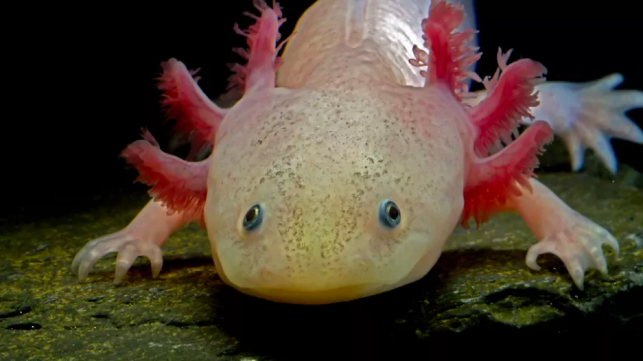
The axolotl (Ambystoma mexicanum) is an aquatic salamander renowned for its ability to regenerate its spinal cord, heart and limbs. These amphibians also readily make new neurons throughout their lives. In 1964, researchers observed that adult axolotls could regenerate parts of their brains, even if a large section was completely removed. But one study found that axolotl brain regeneration has a limited ability to rebuild original tissue structure.
So how perfectly can axolotl‘s regenerate their brains after injury?
As a researcher studying regeneration at the cellular level, I and my colleagues in the Treutlein Lab at ETH Zurich and the Tanaka Lab at the Institute of Molecular Pathology in Vienna wondered whether axolotls are able to regenerate all the different cell types in their brain, including the connections linking one brain region to another. In our recently published study, we created an atlas of the cells that make up a part of the axolotl brain, shedding light on both the way it regenerates and brain evolution across species.
Why look at cells?
Different cell types have different functions. They are able to specialize in certain roles because they each express different genes. Understanding what types of cells are in the brain and what they do helps clarify the overall picture of how the brain works. It also allows researchers to make comparisons across evolution and try to find biological trends across species.
One way to understand which cells are expressing which genes is by using a technique called single-cell RNA sequencing (scRNA-seq). This tool allows researchers to count the number of active genes within each cell of a particular sample. This provides a “snapshot” of the activities each cell was doing when it was collected.
This tool has been instrumental in understanding the types of cells that exist in the brains of animals. Scientists have used scRNA-seq in fish, reptiles, mice and even humans. But one major piece of the brain evolution puzzle has been missing: amphibians.
Mapping the axolotl brain
Our team decided to focus on the telencephalon of the axolotl. In humans, the telencephalon is the largest division of the brain and contains a region called the neocortex, which plays a key role in animal behavior and cognition. Throughout recent evolution, the neocortex has massively grown in size compared with other brain regions. Similarly, the types of cells that make up the telencephalon overall have highly diversified and grown in complexity over time, making this region an intriguing area to study.
We used scRNA-seq to identify the different types of cells that make up the axolotl telencephalon, including different types of neurons and progenitor cells, or cells that can divide into more of themselves or turn into other cell types. We identified what genes are active when progenitor cells become neurons, and found that many pass through an intermediate cell type called neuroblasts—previously unknown to exist in axolotls—before becoming mature neurons.
We then put axolotl regeneration to the test by removing one section of their telencephalon. Using a specialized method of scRNA-seq, we were able to capture and sequence all the new cells at different stages of regeneration, from one to 12 weeks after injury. Ultimately, we found that all cell types that were removed had been completely restored.
We observed that brain regeneration happens in three main phases. The first phase starts with a rapid increase in the number of progenitor cells, and a small fraction of these cells activate a wound-healing process. In phase two, progenitor cells begin to differentiate into neuroblasts. Finally, in phase three, the neuroblasts differentiate into the same types of neurons that were originally lost.
Astonishingly, we also observed that the severed neuronal connections between the removed area and other areas of the brain had been reconnected. This rewiring indicates that the regenerated area had also regained its original function.
Amphibians and human brains
Adding amphibians to the evolutionary puzzle allows researchers to infer how the brain and its cell types has changed over time, as well as the mechanisms behind regeneration.
When we compared our axolotl data with other species, we found that cells in their telencephalon show strong similarity to the mammalian hippocampus, the region of the brain involved in memory formation, and the olfactory cortex, the region of the brain involved in the sense of smell. We even found some similarities in one axolotl cell type to the neocortex, the area of the brain known for perception, thought and spatial reasoning in humans. These similarities indicate that these areas of the brain may be evolutionarily conserved, or stayed comparable over the course of evolution, and that the neocortex of mammals may have an ancestor cell type in the telencephalon of amphibians.
While our study sheds light on the process of brain regeneration, including which genes are involved and how cells ultimately become neurons, we still don’t know what external signals initiate this process. Moreover, we don’t know if the processes we identified are still accessible to animals that evolved later in time, such as mice or humans.
But we’re not solving the brain evolution puzzle alone. The Tosches Lab at Columbia University explored the diversity of cell types in another species of salamander, Pleurodeles waltl, while the Fei lab at the Guangdong Academy of Medical Sciences in China and collaborators at life sciences company BGI explored how cell types are spatially arranged in the axolotl forebrain.
Identifying all the cell types in the axolotl brain also helps pave the way for innovative research in regenerative medicine. The brains of mice and humans have largely lost their capacity to repair or regenerate themselves. Medical interventions for severe brain injury currently focus on drug and stem cell therapies to boost or promote repair. Examining the genes and cell types that allow axolotls to accomplish nearly perfect regeneration may be the key to improve treatments for severe injuries and unlock regeneration potential in humans.
https://medicalxpress.com/news/2022-09-axolotls-regenerate-brains-revealing-secrets.html?utm_content=220147797&utm_medium=social&utm_source=facebook&hss_channel=fbp-479163965435700&fbclid=IwAR2k0MvLaHFhcYlmNFFQeyBA2APO6XEpNnspHS6eh6wZtREsNmvezwMWsvk
Naamgeving
De Nederlandstalige naam axolotl komt uit de Azteekse taal Nahuatl, deze naam wordt geschreven als āxōlōtl. Ook in andere talen wordt deze naam vaak gebruikt. Axolotl heeft verschillende betekenissen zoals waterslaaf of waterhond. In de Spaanse taal wordt de salamander met ajolote aangeduid.
De soortnaam mexicanum verwijst naar het leefgebied van de salamander in Mexico.
Verspreiding en habitat
De axolotl komt alleen voor in Mexico. De salamander komt alleen voor in het midden van het land, ten zuiden van de hoofdstad Mexico-Stad. Het is een bewoner van enkele grote meren, oorspronkelijk kwam de axolotl voor in het Xochimilcomeer in de deelstaat Xochimilco en het Chalcomeer in Midden-Mexico. Vroeger kwam de salamander waarschijnlijk ook voor in de meren Texcoco en Zumpango.
De wilde populaties zijn sterk bedreigd geraakt door de groei van de noordelijker gelegen Mexico-Stad, waardoor het Xochimilcomeer al grotendeels droog is komen te liggen. De met water gevulde delen van het zuidelijke deel van het Xochimilcomeer zijn vandaag de dag de enige bekende natuurlijke habitat van de salamander. Er zijn enkele waarnemingen bekend van de salamander in meren rond Mexico-Stad maar waarschijnlijk zijn deze populaties vertegenwoordigers van de verwante soort Ambystoma velasci.
Habitat
De axolotl komt voor in bergmeren en de aangelegen kanalen. Dergelijke meren liggen op enige hoogte, het Xochimilcomeer is gelegen op een hoogte van meer dan 2200 meter boven zeeniveau. De axolotl is speciaal aangepast op bergmeren en behoudt de juveniele kenmerken zodat het dier permanent onder water kan leven. Er is geen metamorfose zoals bij veel andere salamanders. Een voordeel van het uitblijven van de metamorfose is het op het water aangepaste lichaam van de larve. Volledig getransformeerde molsalamanders hebben een worstvormig lichaam. De poten zijn groter en de staartzomen zijn verdwenen, wat het vermogen tot het zwemmen sterk beperkt. Daarnaast zijn salamanders op het land gevoelig voor uitdroging maar daar heeft de permanent in het water levende axolotl geen last van.
Het leefgebied van de axolotl bestaat uit diepe, permanente wateren in een vulkanische omgeving. Als ze transformeren verliezen ze de niet alleen de visachtige lichaamsvorm maar gaan ook de kieuwen verloren. Voor de axolotl zou dit eveneens alleen maar nadelig zijn. Op het rotsachtige, kale land is weinig voedsel te vinden. Hier staat tegenover dat er in habitats vergelijkbaar met die van de axolotl ook wel soorten molsalamanders zijn gevonden, zoals de tijgersalamander (Ambystoma tigris). https://nl.wikipedia.org/wiki/Axolotl
Regeneration

Dunkles Exemplar
Axolotl verfügen über die Fähigkeit, Gliedmaßen, Organe und sogar Teile des Gehirns und Herzens wiederherzustellen. Die Regenerate sind in der Regel keine Verkrüppelungen, sondern vollständig und funktionstüchtig. Ob das wiederhergestellte neuronale Netzwerk des Gehirns dann auch tatsächlich wie zuvor funktioniert, weiß man aber noch nicht. Nach einer Verwundung bildet sich ein Wundepithel, das auch darunterliegendes Gewebe zur Heilung veranlasst. Nach wenigen Tagen bildet sich bei verlorenen Körperteilen eine Art Regenerationsknospe, aus welcher das Körperteil nachwächst.[11]
Die Regenerationsfähigkeit macht die Art zu einem lohnenden Fo rschungsobjekt] Untersucht werden die Mechanismen, die eine solche Regeneration ermöglichen, z. B. das Enzym LOXe. Bisher nahm man an, dass sich nach einer Verletzung zunächst die umliegenden Zellen in besonders variable, pluripotente Stammzellen zurückentwickeln und im nächsten Schritt aus diesen alle neuen Zellen entstehen. Neuere Forschungen haben ergeben, dass sich Gliedmaßen oder Organe aus Zellen regenerieren, die sich nur jeweils zu bestimmten Gewebetypen weiterentwickeln können. (zu diesem Forschungsansatz siehe auch: Strudelwürmer). Die Regeneration erfolgt in einer zeitlich festgelegten Reihenfolge. Zur Untersuchung der genetischen Ursachen der Regenerationsfähigkeit wurde das Genom des Axolotls sequenziert. Dabei wurde festgestellt, dass dem Axolotl im Gegensatz zu anderen Salamandern das Gen Pax3 fehlt. Dessen Funktion wird von Pax7 übernommen. Mit 32 Milliarden Basenpaaren ist das Genom des Axolotl zehnmal so groß wie das menschliche Genom und damit nach dem Genom des Australischen Lungenfischs das größte, das jemals entschlüsselt wurde. https://de.wikipedia.org/wiki/Axolotl
Use as a model organism

Leucistic axolotl in captivity
Today, the axolotl is still used in research as a model organism, and large numbers are bred in captivity. They are especially easy to breed compared to other salamanders in their family, which are rarely captive-bred due to the demands of terrestrial life. One attractive feature for research is the large and easily manipulated embryo, which allows viewing of the full development of a vertebrate. Axolotls are used in heart defect studies due to the presence of a mutant gene that causes heart failure in embryos. Since the embryos survive almost to hatching with no heart function, the defect is very observable. The axolotl is also considered an ideal animal model for the study of neural tube closure due to the similarities between human and axolotl neural plate and tube formation; the axolotl’s neural tube, unlike the frog’s, is not hidden under a layer of superficial epithelium. There are also mutations affecting other organ systems some of which are not well characterized and others that are. The genetics of the color variants of the axolotl have also been widely studied.
Regeneration
The feature of the axolotl that attracts most attention is its healing ability: the axolotl does not heal by scarring and is capable of the regeneration of entire lost appendages in a period of months, and, in certain cases, more vital structures, such as tail, limb, central nervous system, and tissues of the eye and heart. They can even restore less vital parts of their brains. They can also readily accept transplants from other individuals, including eyes and parts of the brain—restoring these alien organs to full functionality. In some cases, axolotls have been known to repair a damaged limb, as well as regenerating an additional one, ending up with an extra appendage that makes them attractive to pet owners as a novelty. In metamorphosed individuals, however, the ability to regenerate is greatly diminished. The axolotl is therefore used as a model for the development of limbs in vertebrates. There are three basic requirements for regeneration of the limb: the wound epithelium, nerve signaling, and the presence of cells from the different limb axes. A wound epidermis is quickly formed by the cells to cover up the site of the wound. In the following days, the cells of the wound epidermis divide and grow quickly forming a blastema, which means the wound is ready to heal and undergo patterning to form the new limb.
It is believed that during limb generation, axolotls have a different system to regulate their internal macrophage level and suppress inflammation, as scarring prevents proper healing and regeneration. However, this belief has been questioned by other studies. Axolotl’s regenerative properties leave the species as the perfect model to study the process of stem cells and its own neoteny feature. Current research can record specific examples of these regenerative properties through tracking cell fates and behaviors, lineage tracing skin triploid cell grafts, pigmentation imaging, electroporation, tissue clearing and lineage tracing from dye labeling. The newer technologies of germline modification and transgenesis are better suited for live imaging the regenerative processes that occur for axolotls.
Genome
The 32 billion base pair long sequence of the axolotl’s genome was published in 2018 and was the largest animal genome completed at the time. It revealed species-specific genetic pathways that may be responsible for limb regeneration. Although the axolotl genome is about 10 times as large as the human genome, it encodes a similar number of proteins, namely 23,251 (the human genome encodes about 20,000 proteins). The size difference is mostly explained by a large fraction of repetitive sequences, but such repeated elements also contribute to increased median intron sizes (22,759 bp) which are 13, 16 and 25 times that observed in human (1,750 bp), mouse (1,469 bp) and Tibetan frog (906 bp), respectively. https://en.wikipedia.org/wiki/Axolotl
Néoténie et métamorphose
Le terme « néoténie » désigne le fait pour un animal de conserver des caractères normalement juvéniles à l’âge adulte. Dans le cas d’A. mexicanum, il s’agit du maintien des branchies à l’âge adulte, branchies qui disparaissent normalement chez les autres urodèles. Ce maintien découle d’une activité plus faible de sa glande thyroïdienne. Cette adaptation serait une conséquence du climat mexicain d’altitude, le froid défavorisant la forme terrestre qui y est plus exposée.
Un animal exposé artificiellement à l’hormone thyroïdienne va se transformer, perdre ses branchies et acquérir des poumons fonctionnels, ainsi qu’un mode de vie terrestre.
La métamorphose de la larve axolotl en adulte se traduit par l’atrophie de ses branchies, le développement de ses poumons (petits à l’état larvaire), et par une série de transformations de son corps lui permettant de quitter son milieu exclusivement aquatique. Cette métamorphose est très rare dans des conditions naturelles. Elle est parfois provoquée par une baisse radicale du niveau des eaux accompagnée d’un réchauffement important.
En captivité, si l’on déplace un axolotl vivant dans un milieu purement aquatique vers un vivarium dans lequel on réduit progressivement le niveau de l’eau, l’animal menacé d’assèchement cesse d’utiliser ses branchies et privilégie ses poumons. Ainsi, en plusieurs semaines, il se métamorphose petit à petit en adulte.
De façon artificielle, une stimulation chimique de la thyroïde peut déclencher cette transformation, en injectant de l’iode ou une hormone de type thyroxine.
La perte de la capacité à produire eux-mêmes les hormones nécessaires à cette transformation résulte du caractère néoténique de cet animal. De plus, ce changement réduit considérablement l’espérance de vie de l’axolotl, si tant est qu’il y survive. Un axolotl au stade néoténique peut vivre de 10 à 15 ans (mais un individu observé à Paris aurait vécu jusqu’à l’âge de 25 ans). Un individu métamorphosé, lui, ne dépassera guère l’âge de 5 ans. https://fr.wikipedia.org/wiki/Axolotl